Diffuse Scattering CryoEM Data Collection
Accelerating dynamic data collection
We are developing methods to democratize techniques for capturing dynamic conformational ensembles. Our current efforts focus on diffuse scattering and cryogenic electron microscopy (cryoEM).
Diffuse scattering
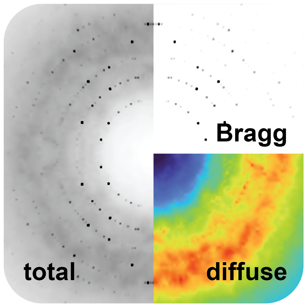
The diffuse signal sits between and beneath the Bragg peaks, encoding how atoms deviate from the average structure.
While diffuse scattering has great promise for detecting protein motion and correlated movement, it is a niche technique used by only a few groups worldwide. But this data is present in essentially all macromolecular diffraction data and has conventionally been discarded as background. X-ray crystallography has always had the potential to detect correlated intra-protein motions. What has been missing is a methodology for collecting, processing, and interpreting the diffuse signal.
The diffuse scattering node of Prism is building the methods, tools, and infrastructure to enable more people to collect, process, and model high-quality diffuse scattering data during every X-ray crystallography experiment.
Improving Data Collection
We are developing protocols to determine how to collect high-quality, reproducible diffuse scattering data. We are creating protocols that can spread across beamlines and groups, enabling reproducibility and interpretability. Our current focus is room-temperature diffraction from highly ordered protein crystals, but we will eventually also understand the diffuse signal in more poorly diffracting samples and across a range of temperatures.
Building Open-source Data Processing Algorithms.
We are building state-of-the-art processing software to process diffuse scattering data. See our software here: mdx2.
Unify Models of Bragg and Diffuse Scattering
Protein motions are currently modeled separately from Bragg peaks and from diffuse scattering, and the two results often disagree. We are developing algorithms based around molecular dynamics and machine learning that learn directly from experimental data, integrating both signals into a single consistent model of the motions that produced them.
CryoEM
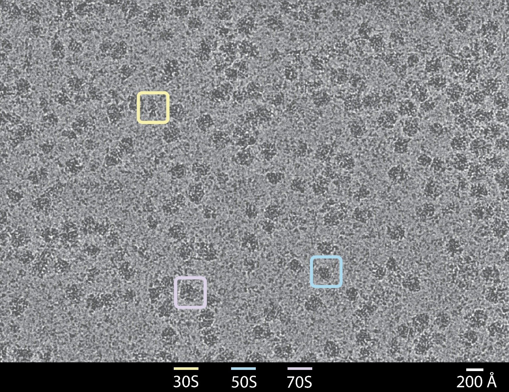
Despite tremendous advances in cryoEM methods over the past decade, the method remains largely focused on recovering the lowest-energy conformational states at high resolution. As an inherently single-molecule technique, cryoEM can capture structural fluctuations spanning the free-energy landscape across a broad range of time and length scales. In some instances, these fluctuations correspond to rare states that are inherently difficult to detect. Such rare states, though transient, can have outsized effects on molecular function.
Using Mutation Perturbations to Identify Conformational Landscape
If many slightly perturbed protein variants were structurally characterized in a coordinated way, each perturbation could act as a probe of the underlying energy surface. Collectively, these probes yield a far richer picture of the landscape, giving direct insight into the regulatory mechanisms that depend on conformational dynamics. Within Prism, we are developing new biochemical methods for sample preparation and advanced image processing approaches to probe this energy landscape at a depth and resolution never before attempted.
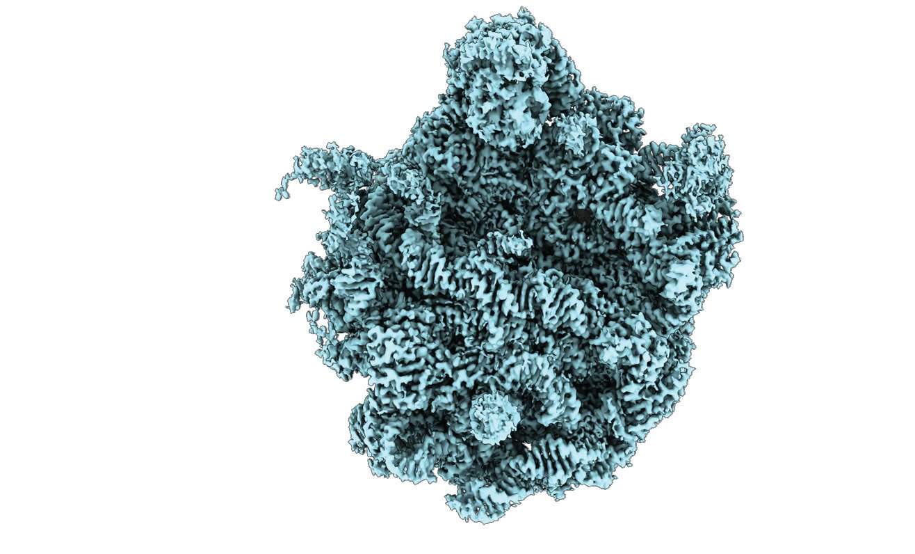
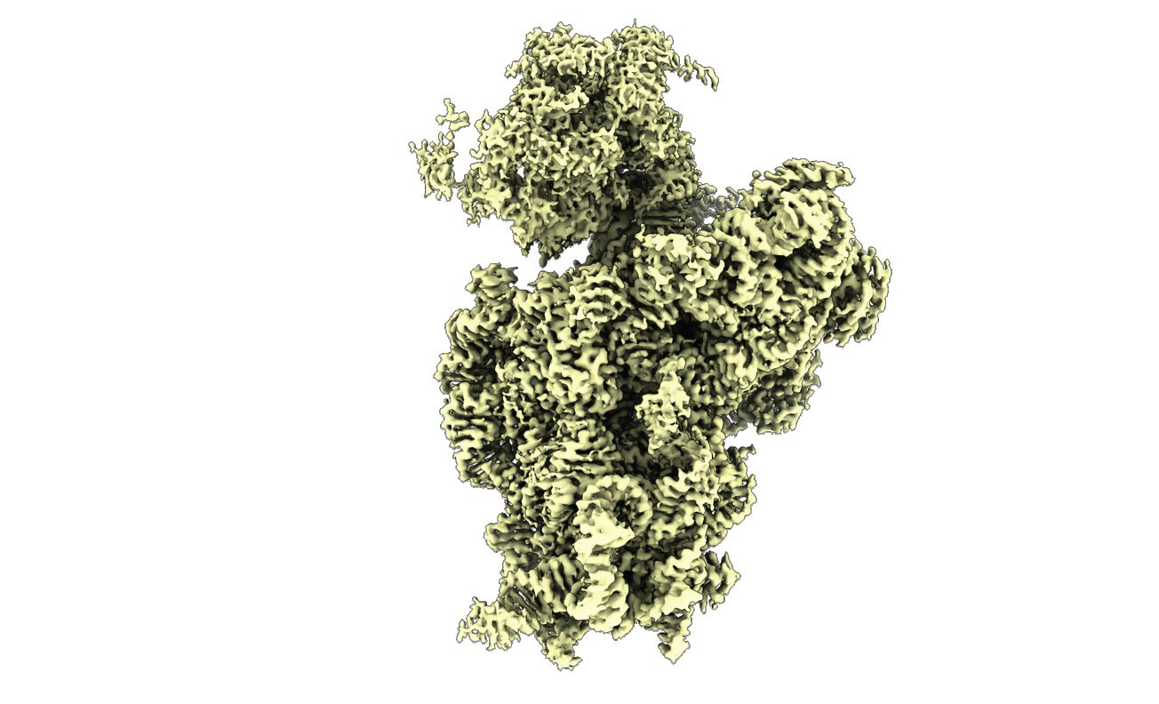
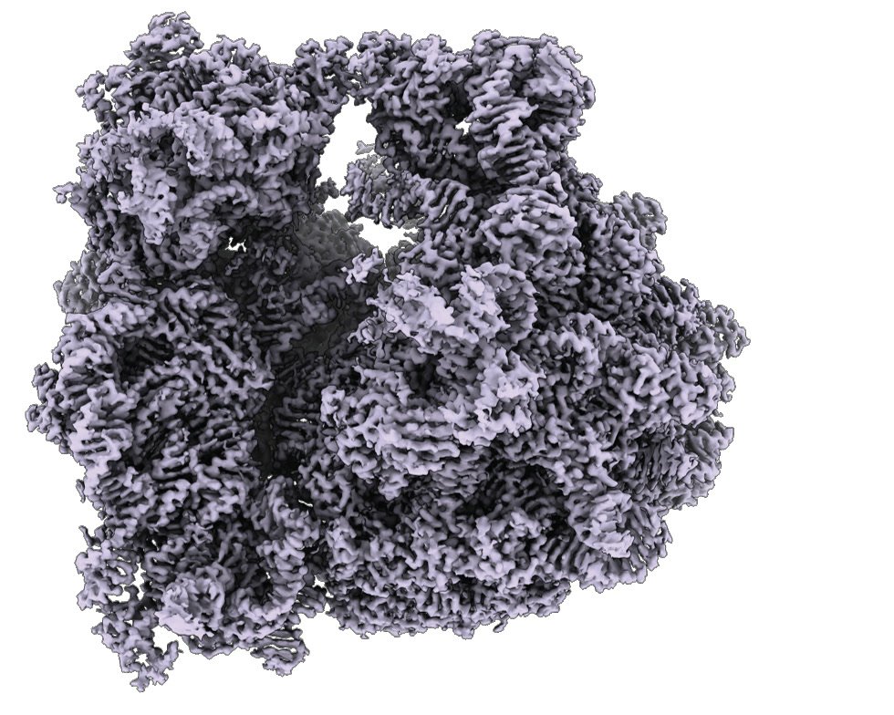
Distinct conformational states recovered from the same pooled experiment.
Increasing Throughput and Target Range in Data Collection
As a single-particle method that images samples under near-native conditions and tolerates inhomogeneity, cryoEM offers strong inherent advantages for assessing structural energy landscapes. Its central limitation is that is has historically been limited to imaging relatively large macromolecular complexes (> 100 kDa). To extend the range of cryoEM, and to align the molecular targets we aim to analyze with those accessible by X-ray crystallography, we are designing ‘scaffolding proteins’ to display our small structural targets ( < 50 kDa) in a manner amenable to structure determination by cryoEM. This approach also involves developing purpose-built data-processing pipelines that leverage constraints from the scaffold protein to expand the molecular-weight range accessible by cryoEM.
Computational Modeling of Ensembles
No existing algorithm adequately handles the map-to-model problem for conformationally heterogeneous data, particularly when states overlap between perturbations or when distinct states within a perturbation are structurally similar. Further, translating these changes to well-encoded models will help close the gap that currently makes heterogeneity analysis a bottleneck.