Diffuse Scattering Modeling Blog
Don't just do something, sit there: Mindful of the MD Model
2 min read
Words by:

Michael WallSenior Scientist – Los Alamos National Laboratory
Recently Taylor Womack wrote about combining GOODVIBES lattice vibrations with MD simulations to improve models of diffuse scattering. Our efforts have begun with MD simulations of P1 lysozyme, for which accurate vibrational models of diffuse scattering are already available.
Our first simulations were prepared using PDB ID 4LZT, which was published in 1997, and was used for previous crystalline MD simulations of diffuse scattering. As is typical, we assessed the MD model by performing an initial simulation where heavy atoms are restrained to their positions in the crystal structure, and compared the mean electron density to the crystal structure. The model was generally reasonable, but the agreement wasn’t as good as we’re used to seeing. In particular, the density appeared to violate the crystal symmetry (Fig. 1).
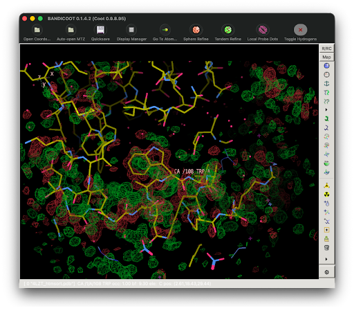
Figure 1. Simulated supercell minus mean unit cell difference density for 4LZT. Green is positive, red is negative. The view is in the neighborhood of Trp108 in one out of 125 unit cells in the 5x5x5 system. The differences are large, indicating inconsistency with the crystal symmetry.
Given these issues, we developed an MD model from PDB ID 6O2H, which was the structure deposited from the diffuse scattering study. The mean density from the initial restrained simulation of 6O2H appeared to agree better with the crystal structure, and to be more consistent with the crystal symmetry (Fig. 2). A quantitative analysis (not shown) supports these qualitative observations.
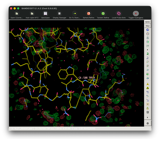
Figure 2. Simulated supercell minus mean unit cell difference density for 6O2H, same view and contour level as Figure 1. Green is positive, red is negative. The differences are smaller overall than for 4LZT, and are largely confined to non-protein regions, whereas the 4LZT differences extend onto the protein itself.
Our experience with P1 lysozyme indicates that, even when MD simulations are restrained to the crystal structure, proteins can “shift in their seats” depending on their location in the crystal. We have previously seen that analysis of local water and protein density features can suggest ways of improving crystalline MD models. Analysis of crystal symmetry in MD simulations might be another useful approach.
Analysis of the P1 lysozyme simulations is ongoing, stay tuned…
Share article: